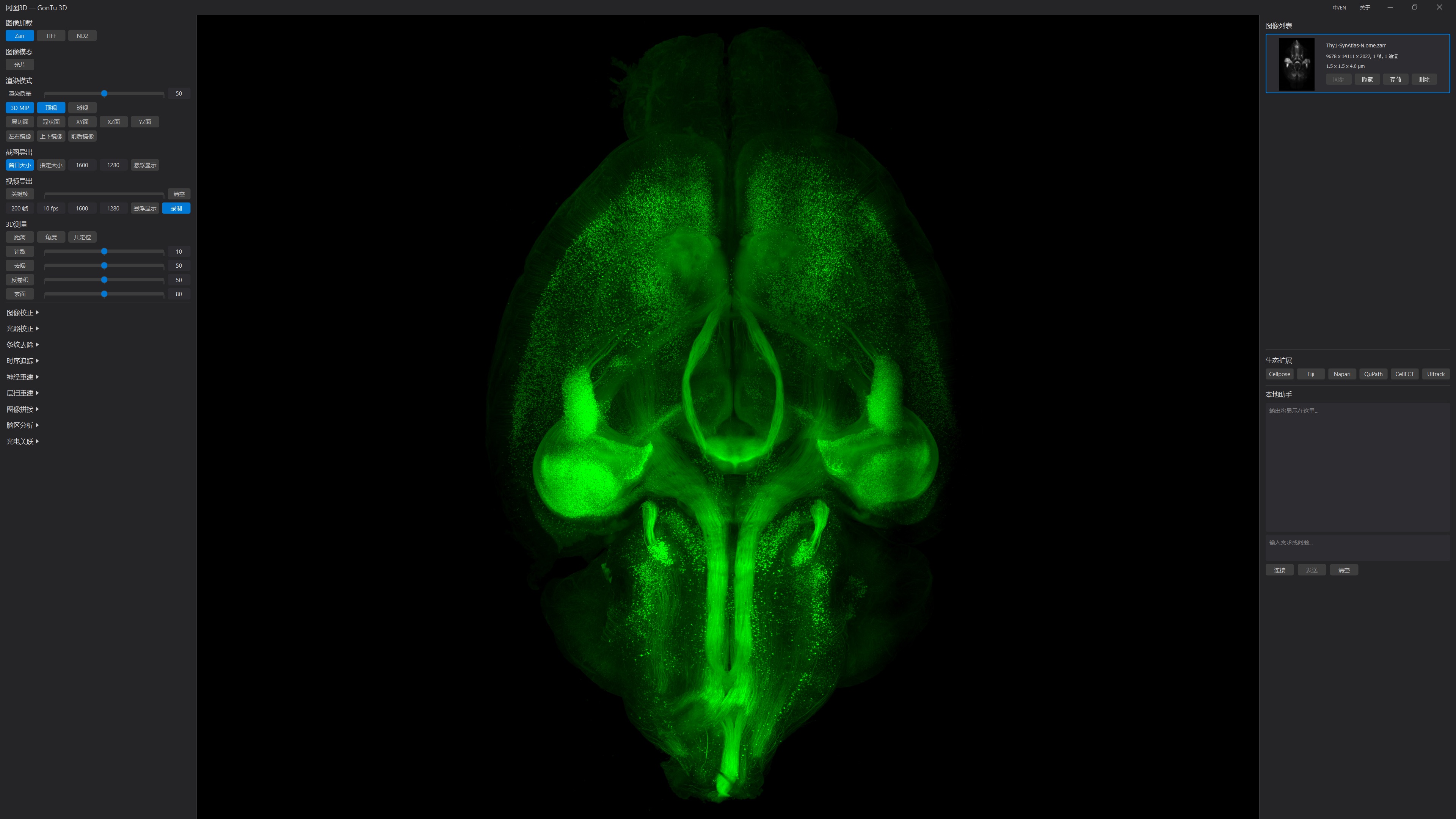
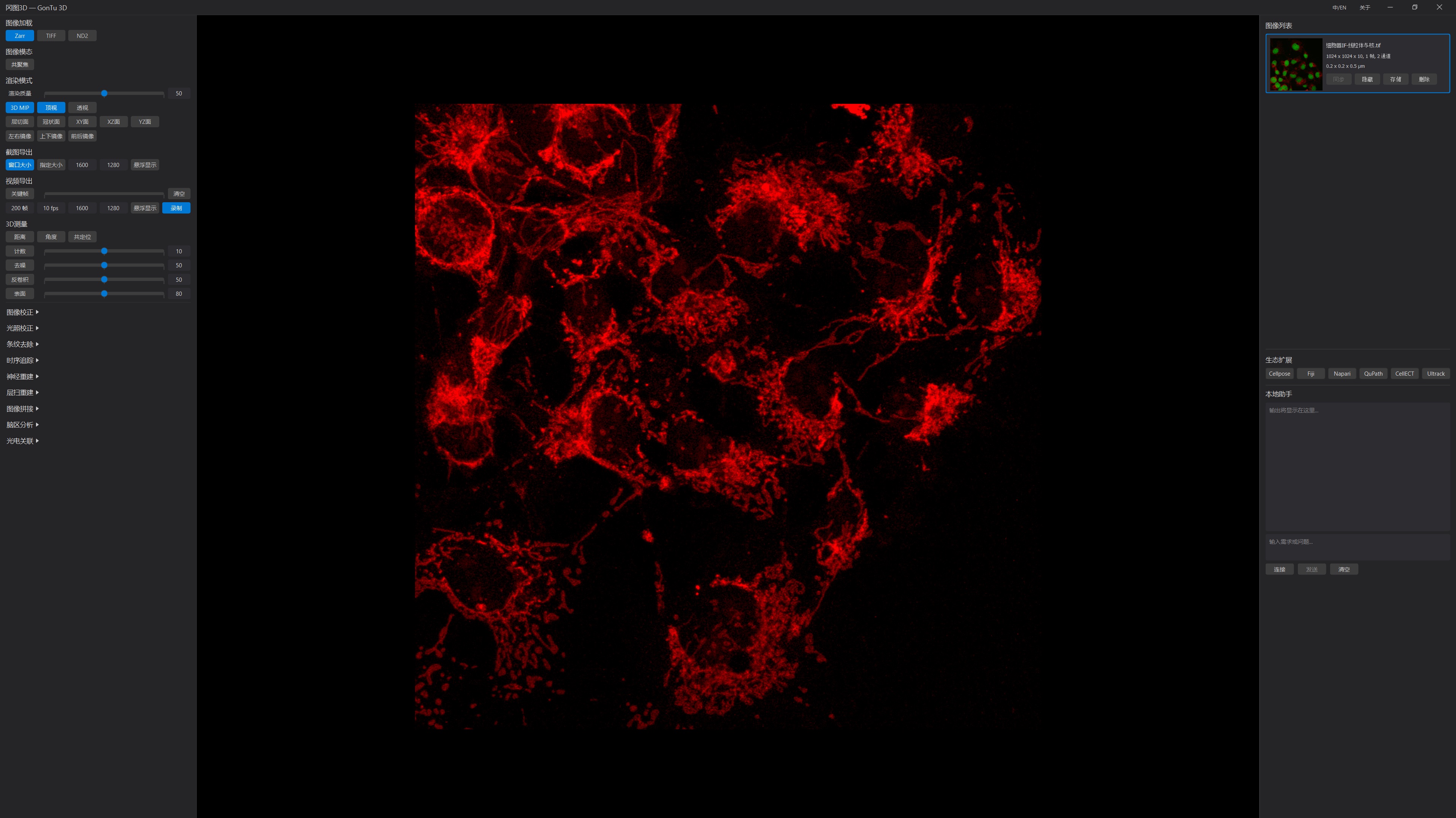
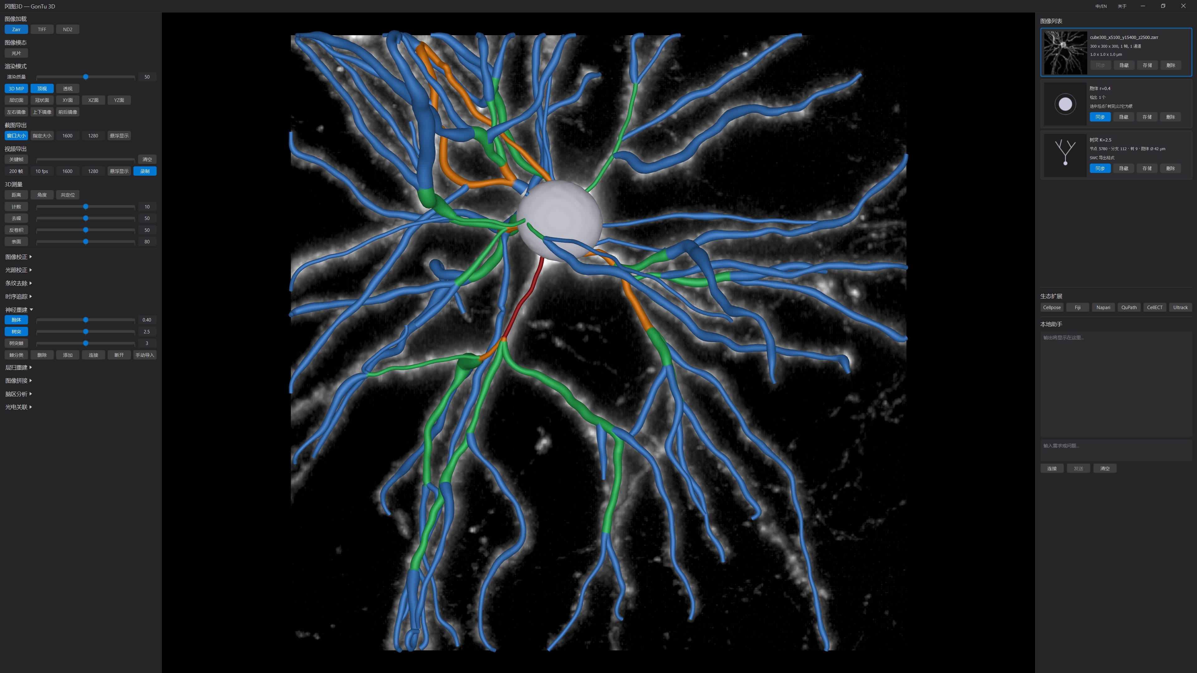
Sample courtesy: Dr. Qinghong Shan, The First Affiliated Hospital of Anhui Medical University
Microscopy image processing software — rendering, correction, and analysis for TB-scale data
Delivered with every license; optional modules combine on top of it.
OME-Zarr, TIFF, and ND2 load natively; for large datasets, first convert to OME-Zarr with the free GonTu Converter for smoother loading and rendering.
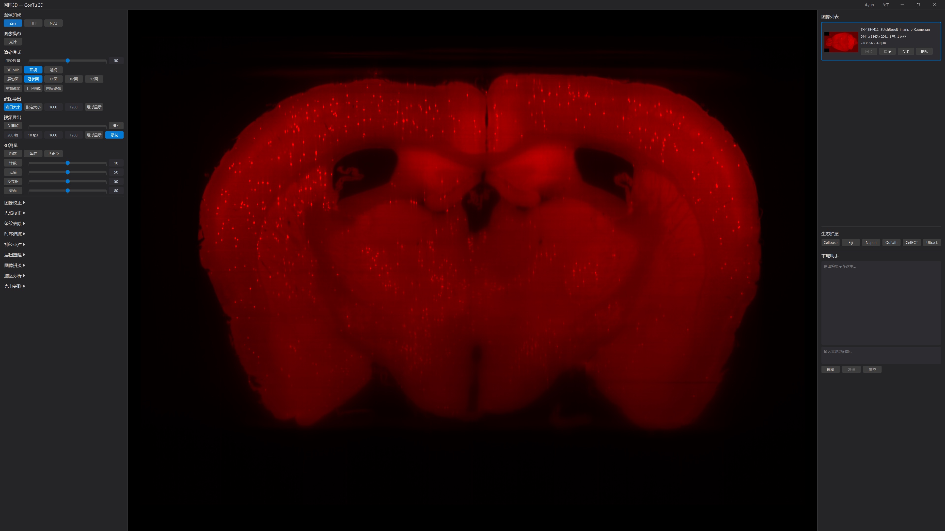
Volume rendering and slicing switch in place at the same angle and zoom; one-click coronal view.
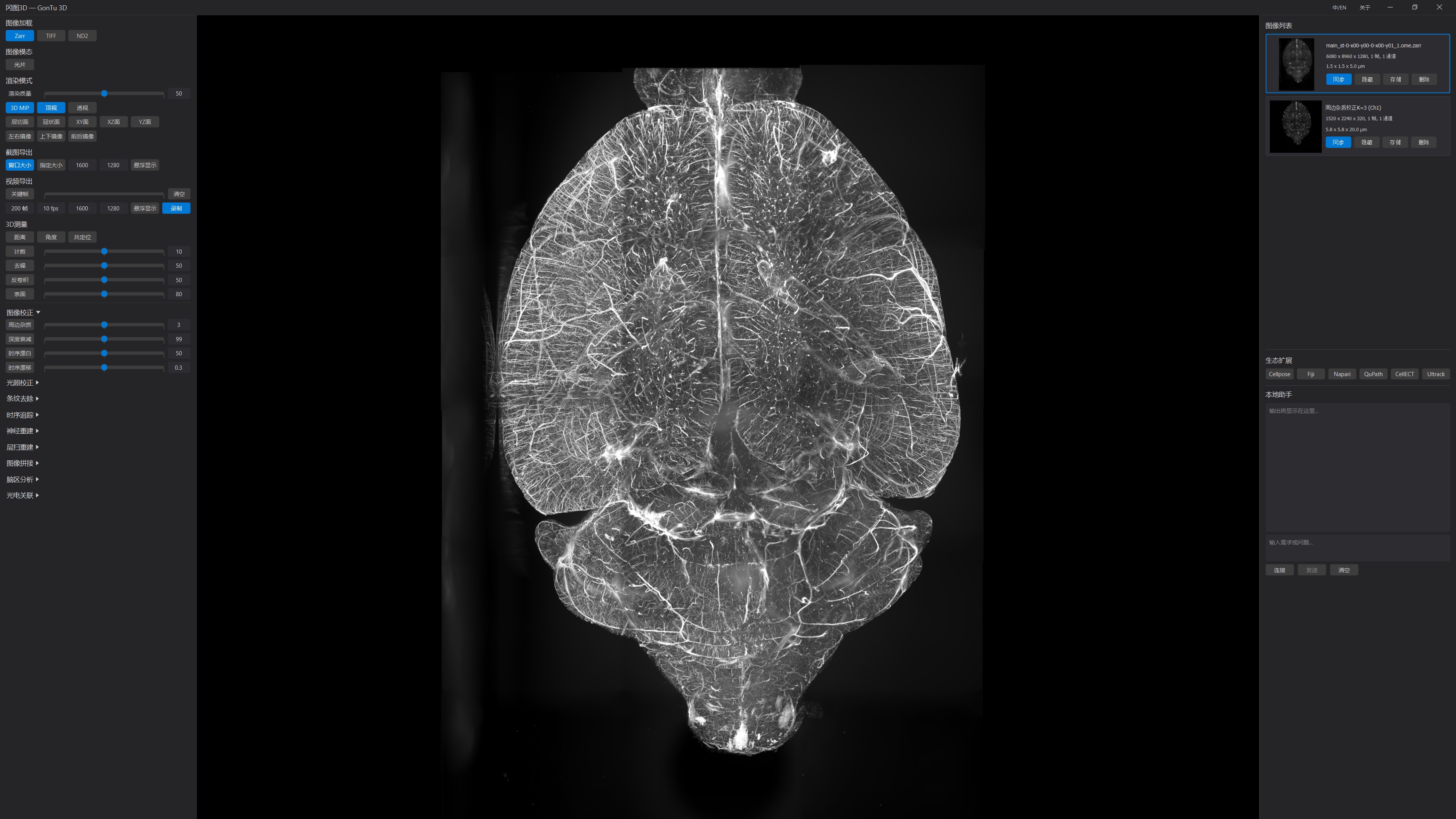
A 16 GB laptop (SSD required) browses TB-scale 3D data smoothly.
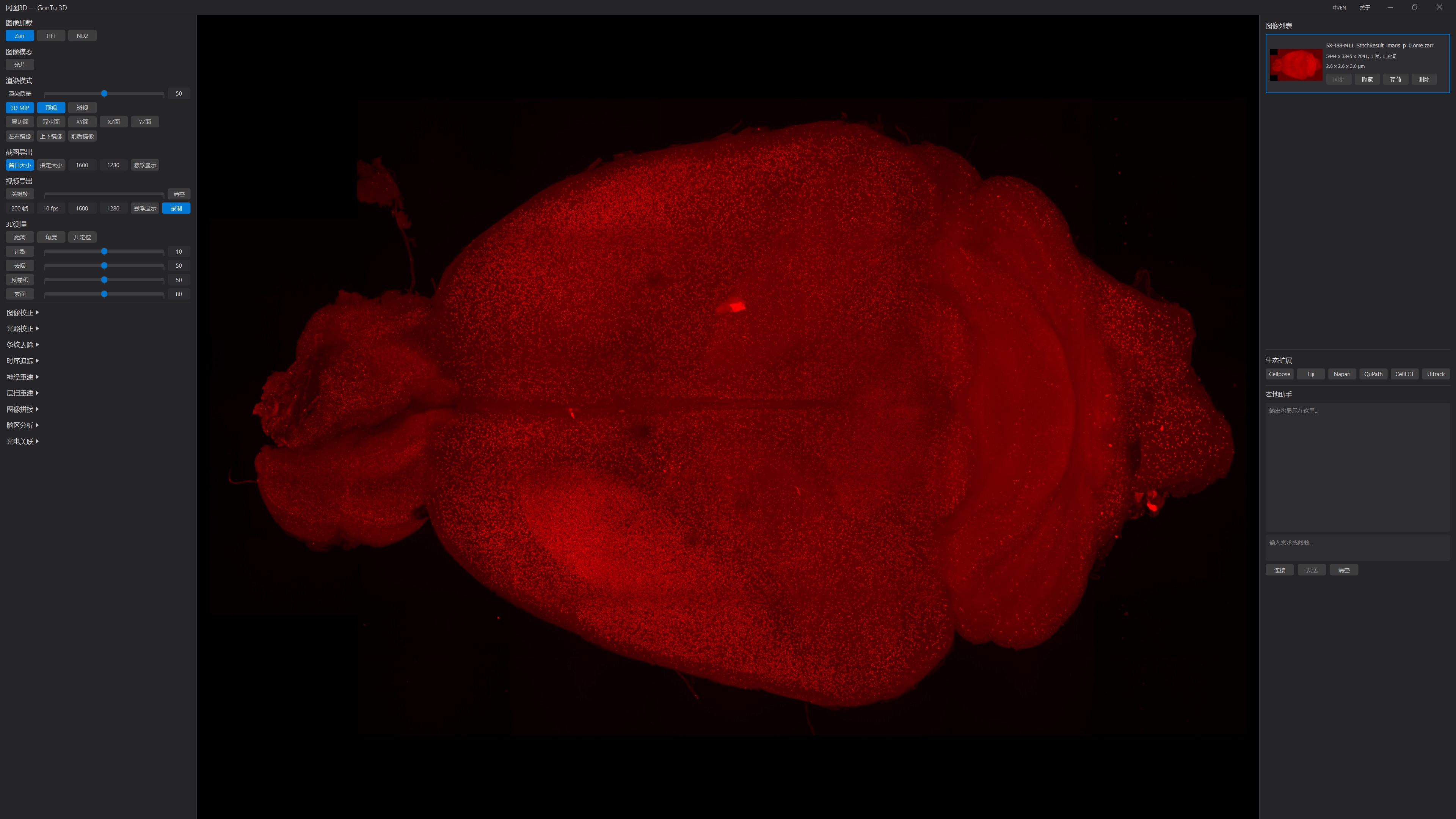
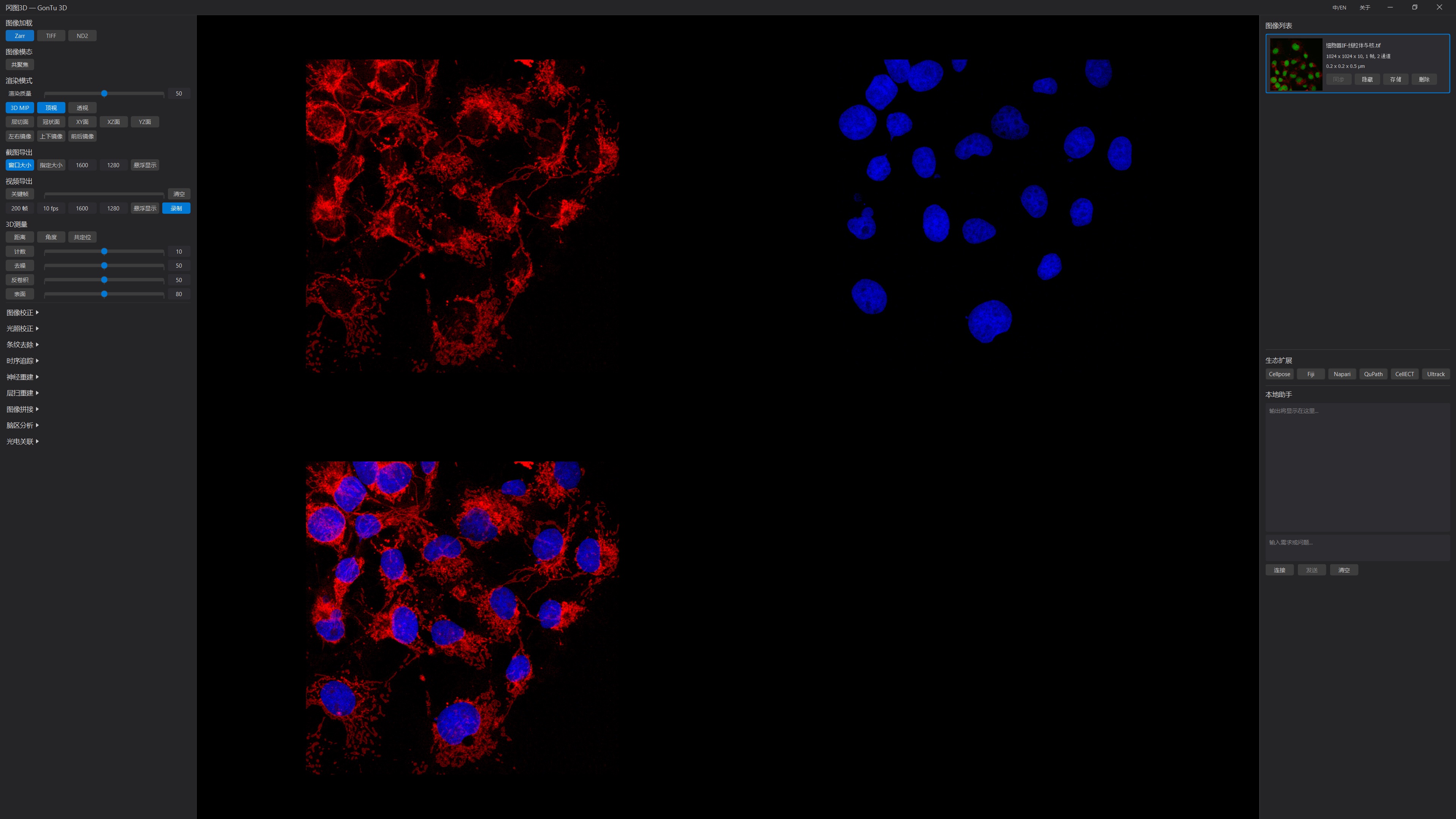
Multiple single channels and fused channels display in sync; double-click to switch between the tiled multi-channel view and single-channel view.
High-resolution RGB TIF snapshots and keyframe-interpolated MP4 export — publication-ready figures in one click.
Distance, angle, and colocalization; cell counting; surface reconstruction and quantification (volume / surface area, STL export); deep-learning denoising and deconvolution; results as cards, one-click CSV export.
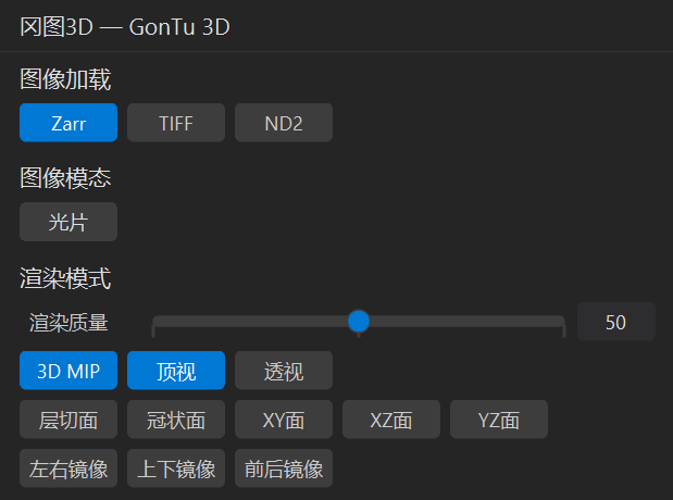
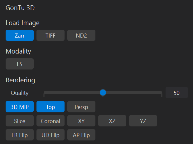
Switch interface language instantly, with no restart; the Chinese UI is deeply adapted, not an afterthought translation.
Configured on demand — independently licensed, freely combined.
Debris removal, depth attenuation / bleaching / drift correction — combinable into a correction chain replayed at full resolution.
Tile-level brightness equalization for stitched data; anomalous tiles detected automatically, corrections feathered seamlessly.
Object tracking with trajectory management.
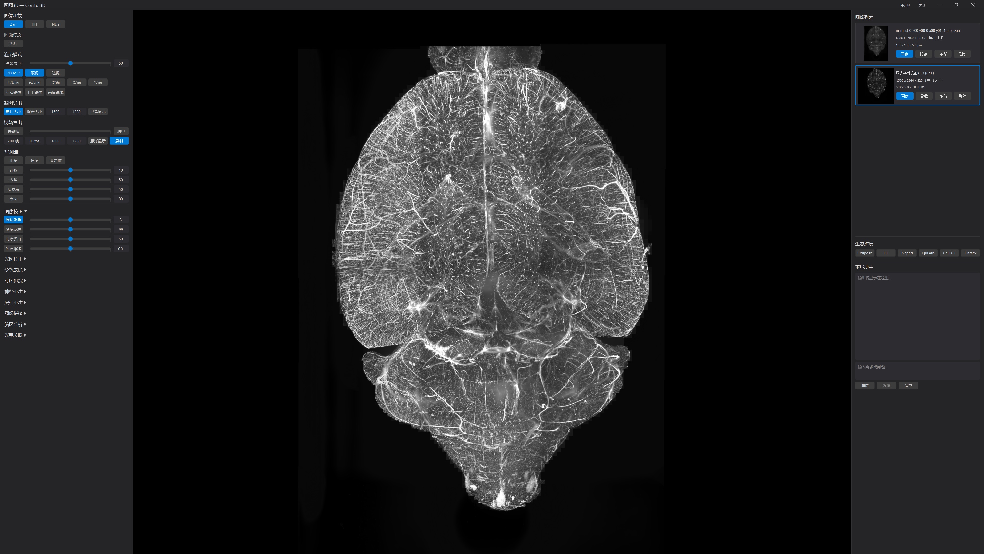
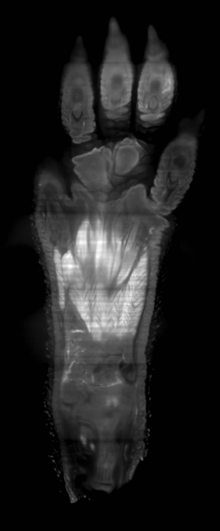
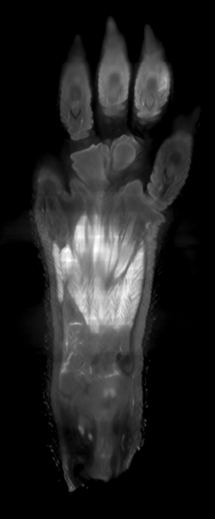
Deep-learning stripe removal for horizontal, vertical, and both directions; single-layer training with full-volume inference; the UI never freezes during processing.
18 built-in scan patterns, sub-pixel registration, giant-image output.
Automatic alignment and stacking of serial sections with global adjustment.
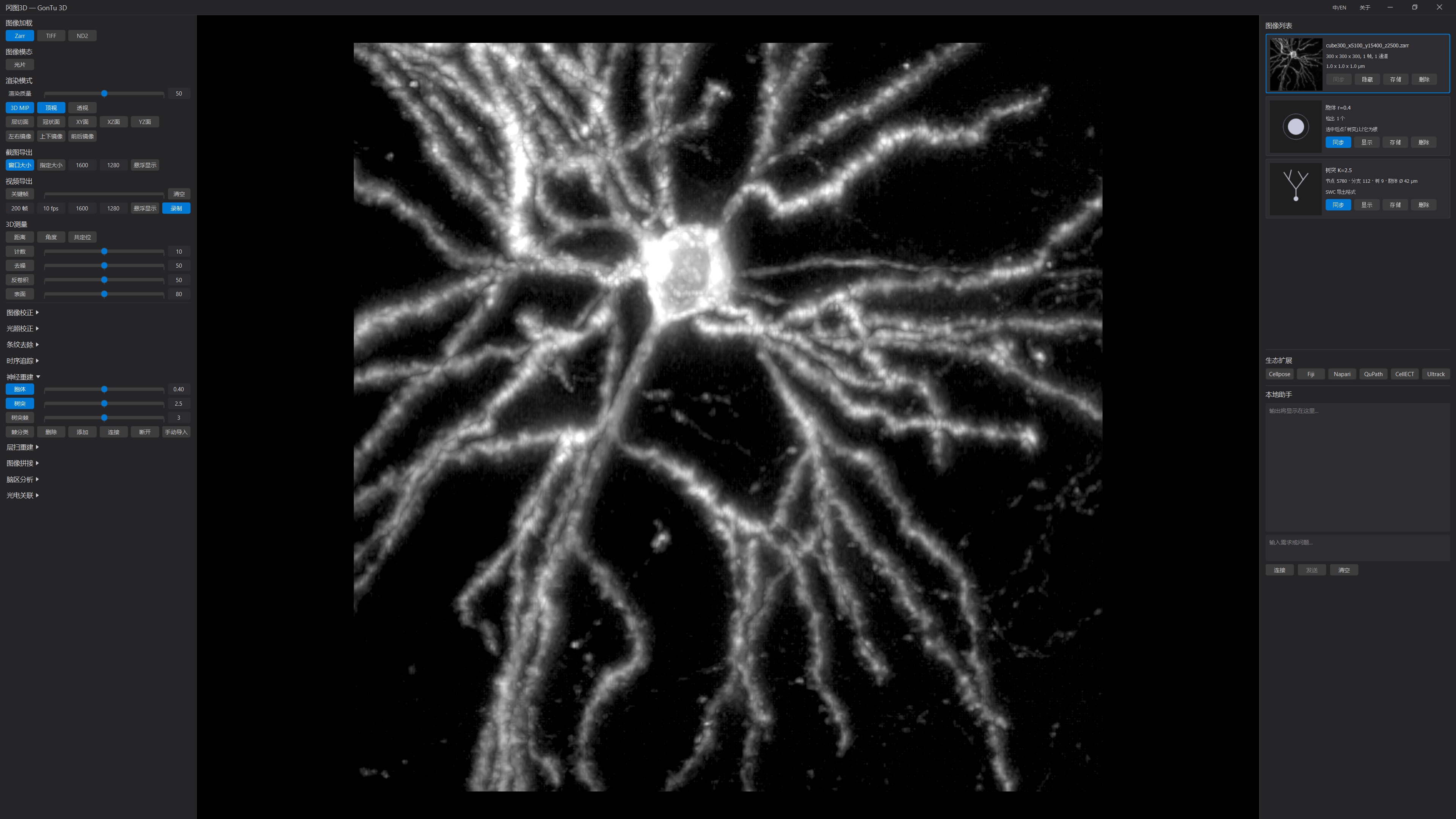
One-click automatic 3D neuron reconstruction with SWC output; spine detection; Strahler-graded coloring.
Correlative light-electron microscopy registration up to 9 degrees of freedom, with baked export.
Standard data interchange with Cellpose, Fiji, Napari, QuPath, and other open-source tools (their copyright remains with their developers; installation required).
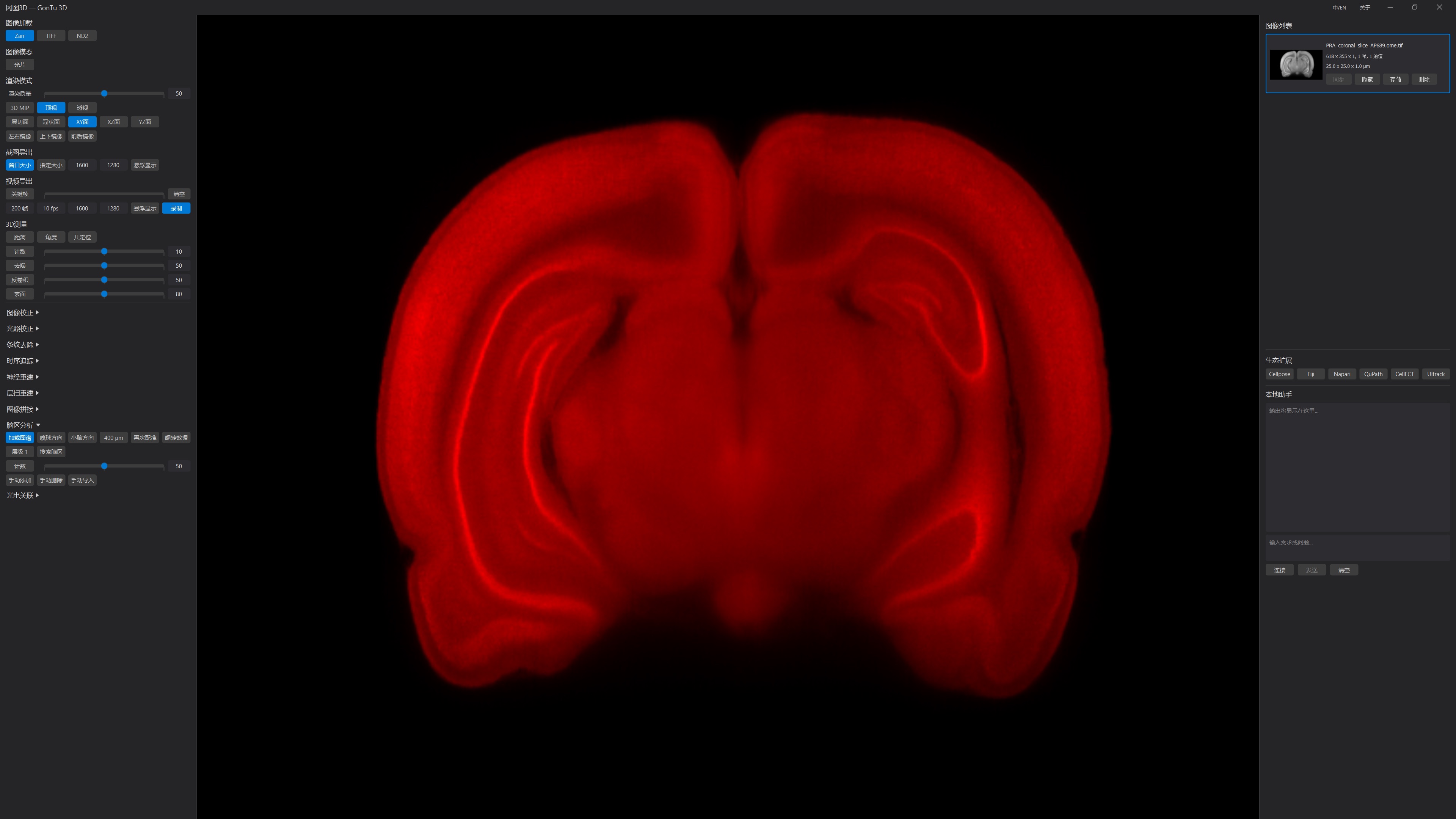
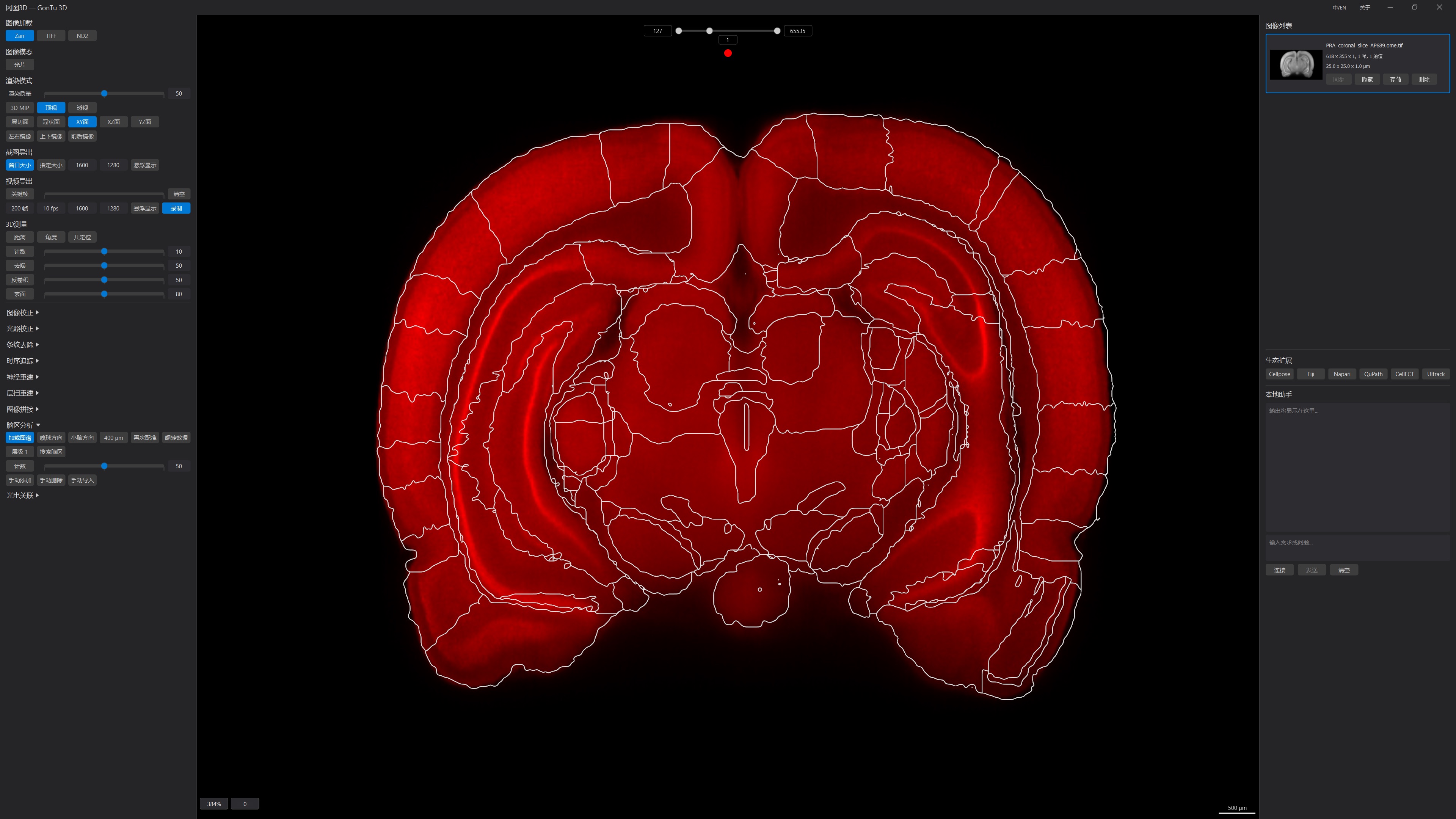
Automatic atlas registration for mouse, rat, and zebrafish brains, with per-region quantification exported to CSV; compatible with major atlases such as Allen CCF and Waxholm Space — atlas files are supplied and loaded by the user.
Local large-language-model assistant — fully offline, data never leaves your machine.
Detailed technical materials available on request.
| Operating system | Windows 10/11 (64-bit) |
|---|---|
| Supported formats | OME-Zarr / TIFF / ND2 (native) |
| Rendering | Supports TB-scale data; full-resolution streaming, no full-volume loading |
| Minimum memory | 16 GB (for TB-scale 3D browsing) |
| UI languages | Chinese / English, hot-switchable |
| 3D measurement | Included in base platform: distance / angle / colocalization / cell counting / surface reconstruction & quantification (STL export) / denoising / deconvolution, CSV export |
| Image correction (optional) | Debris removal, depth attenuation / bleaching / drift correction |
| Illumination correction (optional) | Mosaic brightness equalization, automatic outlier-tile detection |
| Destriping (optional) | Deep-learning stripe removal (horizontal / vertical / bidirectional) |
| Time-series tracking (optional) | Object tracking and trajectory management |
| Neuron reconstruction (optional) | One-click automatic reconstruction, standard SWC output; spine detection to CSV |
| Serial-section reconstruction (optional) | Automatic alignment and stacking of serial sections into 3D volumes |
| Image stitching (optional) | 18 built-in scan patterns, sub-pixel registration |
| Brain-region analysis (optional) | Mouse / rat / zebrafish atlas registration, per-region quantitative statistics |
| CLEM (optional) | Registration up to 9 degrees of freedom |
The elegance, intelligence, and ease of use of GonTu 3D make image processing simple and efficient.